
Fig. 6. The knockout of Zcwpw1 leads to POI.
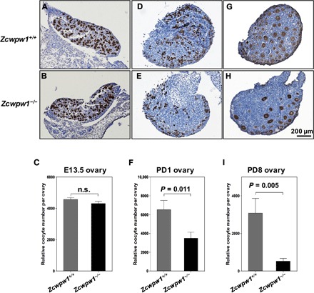
(A and B) Representative Zcwpw1+/+ (A) and Zcwpw1−/− (B) ovary sections from E13.5 females immunostained for mouse vasa homolog (MVH) with hematoxylin counterstaining. (C) Oocyte counts (relative numbers) showed that there were similar numbers of oocytes in E13.5 Zcwpw1+/+ and Zcwpw1−/− females. MVH-positive cells were counted. (D and E) Representative Zcwpw1+/+ (D) and Zcwpw1−/− (E) ovary sections from PD1 females immunostained for MVH with hematoxylin counterstaining. (F) Relative oocyte counts showed that Zcwpw1−/− ovaries contained significantly fewer oocytes than Zcwpw1+/+ ovaries. (G and H) Representative Zcwpw1+/+ (G) and Zcwpw1−/− (H) ovary sections from PD8 females immunostained for MVH with hematoxylin counterstaining. (I) Relative oocyte counts showed that Zcwpw1−/− ovaries contained significantly fewer follicles than Zcwpw1+/+ ovaries. MVH-positive oocyte nuclei with characteristic surrounding granulosa cell layers were counted. In all cases, counts were made for every section (8 μm per section) and summed to calculate the total number of oocytes per ovary. For each genotype, six ovaries from three mice were analyzed. P values were calculated by Student’s t test.