
Figure 3.
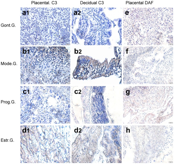
(e) Immunohistochemical staining method was used to detect decay accelerating factor (DAF) (e–h) expression and C3 (a1–d1, a2–d2) deposition in placenta on day 15·5 of pregnancy. Placenta sections of control group (Cont.G.) (a1, a2, e), model group (Mode.G.) (b1, b2, f), progesterone group (Prog.G.) (c1, c2, g) and oestrogen group (Estr.G.) (d1, d2, h) were incubated with goat anti‐mouse decay accelerating factor (DAF) monoclonal antibody (mAb) or goat anti‐mouse C3/C3a mAb, followed by rabbit anti‐mouse immunoglobulin (Ig)G conjugated to horseradish peroxidase (HRP) and detected by diaminobenzidine (DAB) and were counterstained with haematoxylin. Positive sites were stained brown. Results are a representation of experiments performed on at least three independent individual samples (×40 magnification).