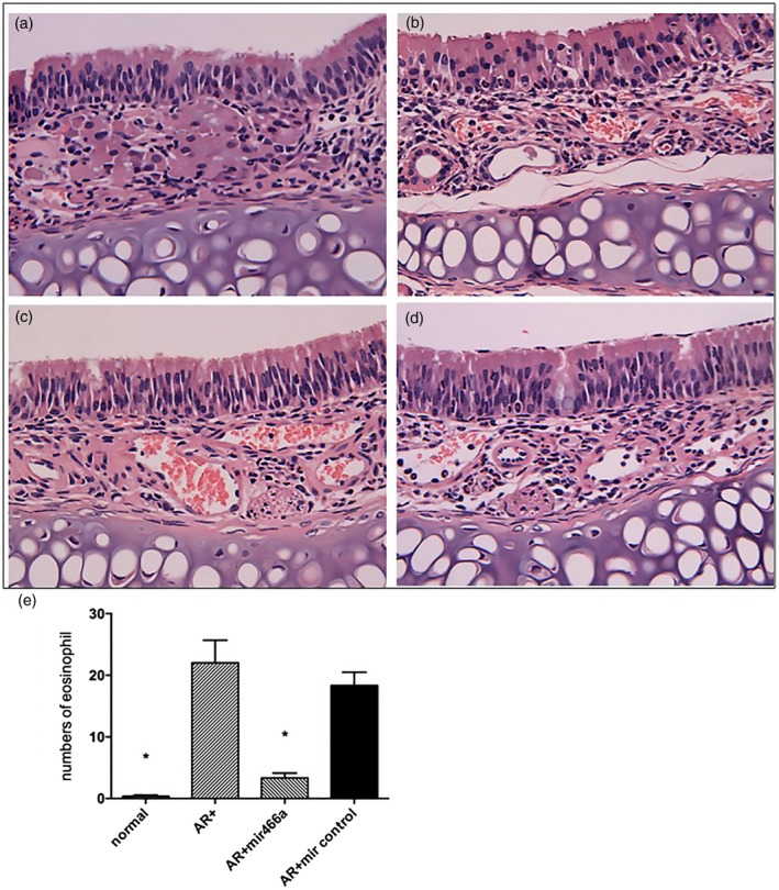
Figure 7.

Histological analysis of nasal mucosa in the (a) normal group, (b) allergic rhinitis (AR) group, (c) AR + lenti‐miR‐466a‐3p group and (d) AR + lenti‐miR control group (magnification ×400). The normal group demonstrated no inflammatory changes, while the nasal mucosa was infiltrated with eosinophils in the AR group and the AR + lenti‐miR control group (white arrow). Eosinophilic infiltration was markedly reduced in the AR + lenti‐miR‐466a‐3p group compared to the AR group. (e) The number of eosinophils in the four groups (per ×400 microscopic field).
