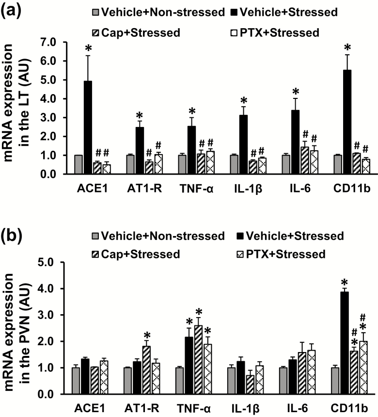
Figure 2.
Quantitative comparison of the mRNA expression of renin–angiotensin system components, proinflammatory cytokines, and microglial marker in the lamina terminalis (LT) and paraventricular nucleus (PVN) in nonstressed, stressed, stressed with pretreatment with either captopril (Cap) or pentoxifylline (PTX) rats before angiotensin II infusion (a and b) (n = 5 per group; *P < 0.05 vs. nonstressed rats; #P < 0.05 vs. stressed rats without pretreatment).

