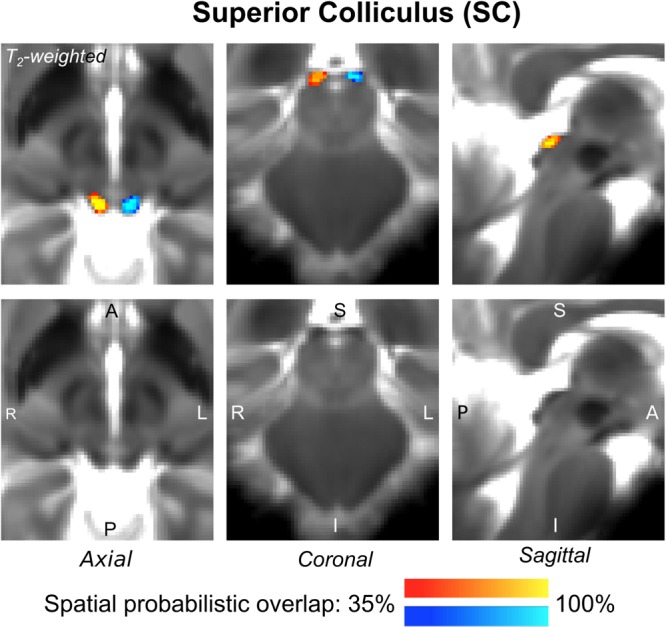
FIGURE 2.

Probabilistic atlas label in MNI space of the SC (left: blue-to-cyan; right: red-to-yellow). The SC was hypointense in T2-weighted MRI; its label is overlaid on the group average T2-weighted image. Very good (i.e., up to 100%) spatial agreement of labels across subjects was observed indicating the feasibility of delineating the probabilistic label of this nucleus involved in visual and oculo-motor functions.
