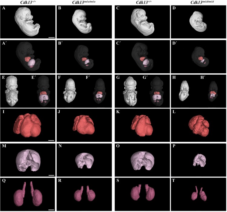
FIGURE 11.
Comparison of Cdk13tm1a and Cdk13tm1d mice at E13.5 High-contrast differentiation resolution by X-ray computed microtomography where Lugol’s staining was used to visualize the soft tissues. The Cdk13tm1a/tm1a, Cdk13tm1d/tm1d and their littermate controls were analyzed at E13.5. (A–D) Overall view of Cdk13+/+, Cdk13tm1a/tm1a, and Cdk13tm1d/tm1d embryos. Both mutants exhibited smaller body size, with severe growth retardation in Cdk13tm1d/tm1d animals. (A’–D’) 3D reconstruction of heart (red color), kidney (violet color) and liver (light pink color) in right side view of embryo, with embryo outlined in gray where segmentation of serial sections was used for the reconstruction. Liver in Cdk13tm1a/tm1a and Cdk13tm1d/tm1d embryos were smaller than in Cdk13+/+ embryos. (E–H’) Frontal view of embryos with 3D reconstruction of heart, kidneys and liver. Clear deficiency in development of midfacial structures in Cdk13tm1d/tm1d embryos in contrast to Cdk13+/+ animals was observed. (I–L) 3D reconstruction of heart. The heart of Cdk13tm1a/tm1a and Cdk13tm1d/tm1d embryos were smaller in comparison to control mice with severe ventricle deficiency detected in Cdk13tm1d/tm1d animals. (M–P) 3D reconstruction of liver. Generally smaller liver were observed in Cdk13tm1a/tm1a and Cdk13tm1d/tm1d embryos in comparison to Cdk13+/+ littermate control mice. Liver in Cdk13tm1d/tm1d mice exhibit abrogated liver parts arrangements. (Q–T) 3D reconstruction of kidneys. Kidneys are smaller with abnormal shape in Cdk13tm1a/tm1a and Cdk13tm1d/tm1d embryos. Scale bar (A–H) = 1,5 mm, Scale bar (I–L) = 350 μm, scale bar (M,P) = 500 μm, and scale bar (Q–T) = 250 μm.

