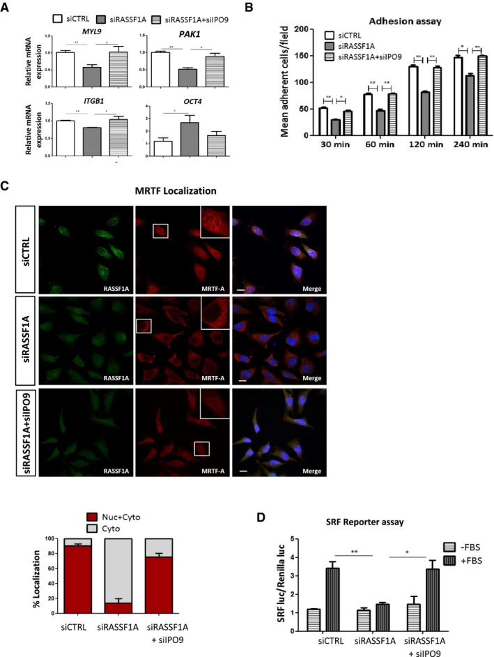
Figure 5. Loss of RASSF1A expression alters MRTF‐A/SRF axis.

- qRT–PCR validation of selected genes known to be affected by the levels of nuclear actin. Transcript levels of MYL9, ITGB1, PAK1 and OCT4 from HeLa cells treated either with siRASSF1A or with siRASSF1A + siIPO9 are relative to GAPDH and normalised to siCTRL cells. Data represent SEM of three independent experiments.
- Adhesion assay. siRNA against RASSF1A significantly decreased HeLa cells’ adhesive rate at all the determined time points compared with control. The cells were cultured for 48 h before harvesting and reseeding for 1 h on 96‐well plates coated with FN. Data represent the SEM of three independent experiments.
- Representative immunofluorescence images of MRTF‐A localisation in siRASSF1A and siRASSF1A + siIPO9. Lower: the MRTF‐A localisation was scored as nuclear/cytoplasmic or predominantly cytoplasmic in 100‐200 cells. DNA was stained with DAPI. Error bars derive from two independent experiments and represent the SEM. Scale bars = 10 μm.
- Luciferase assay of SRF‐dependent promoter in cells transfected with siCTRL, siRASSF1A or siRASSF1A + siIPO9 following stimulation with 10% FBS for 5 h. Data are expressed as SRF luciferase activity relative to Renilla control and represent the SEM of two independent experiments.
