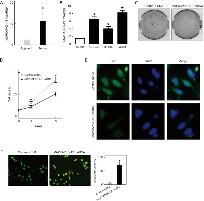
Figure 5.
MAPKAPK5-AS1 could regulate the proliferation of LUAD cell. (A) MAPKAPK5-AS1 was over-expressed in LUAD patient tissue samples; *P<0.05 vs. adjacent tissues. (B) MAPKAPK5-AS1 was up-regulated in LUAD cell lines. 16HBE served as control cells; *P<0.05 vs. control. (C) Silencing of MAPKAPK5-AS1 decreased colony formation capability. A549 cells were seeded and transfected with MAPKAPK5-AS1 siRNA or control siRNA, respectively. The colony number was assessed by crystal violet staining; *P<0.05 vs. control (n=3 independent experiments). (D) MAPKAPK5-AS1 knockdown resulted in growth inhibition of A549 cells. A549 cells transfected with MAPKAPK5-AS1 siRNA or control siRNA were seeded in 96-well plates for 48 h culturing, and cell viability was analyzed by a CCK-8 assay; *P<0.05 vs. control (n=3 independent experiments). (E) Silencing of MAPKAPK5-AS1 suppressed A549 cell proliferation. A549 cells were seeded and transfected with MAPKAPK5-AS1 siRNA or control siRNA. Immunofluorescence staining of Ki-67 was performed to analyze the cell proliferation capability. (F) Silencing of MAPKAPK5-AS1 induced apoptosis in LUAD cells. A549 cells were seeded and transfected with MAPKAPK5-AS1 siRNA or control siRNA for 48 h. Cells were fixed and subjected to AO/EB staining to detect changes in their nuclei. The orange-colored region indicates the initiation of apoptosis; *P<0.05 vs. control (n=3 independent experiments). LUAD, lung adenocarcinoma.

