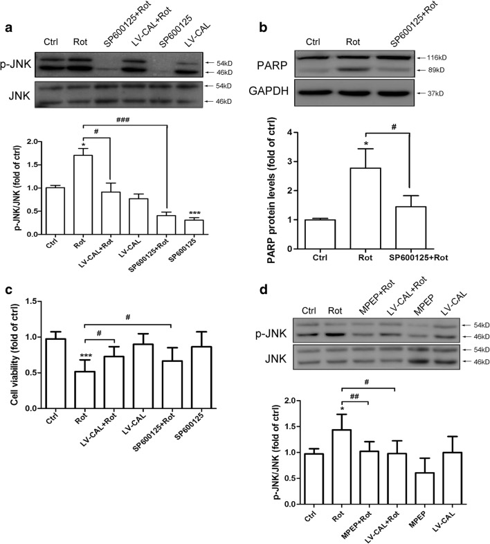
Fig. 5.
CAL suppresses mGluR5-mediated JNK phosphorylation and ameliorates MN9D cell apoptosis induced by rotenone administration. (A) Cells with LV-CAL infection or SP600125 pretreatment were serum-starved for 16 h followed by rotenone treatment (0.2 μM) for 1 h, and then harvested for p-JNK analysis (up). The protein level was normalized to total JNK and represented as the fold difference of the control group (down). (B) Cells were pretreated with SP600125 (100 μM, 30 min) followed by rotenone (0.2 μM) for 24 h and then harvested for PARP detection by Western blotting (up). The protein level was normalized to GAPDH and represented as the fold difference of the control group (down). (C) Cells were infected with LV-CAL or pretreated with SP600125 (100 μM, 30 min) followed by rotenone (0.2 μM) treatment for 24 h, and then cell viability was estimated by the MTT assay. (D) Cells were pretreated with MPEP (100 μM, 30 min) or infected with LV-CAL followed by rotenone (0.2 μM) for 1 h, then cell lysates were collected for p-JNK assessment (up). The protein level was normalized to total JNK and represented as the fold difference of the control group (down). Untreated cells were used as controls. Data shown in all panels of this figure represent the mean ± SEM of 3 independent experiments. The statistical significance was determined by 1-way ANOVA followed by Dunnett’s test. *p < 0.05 and ***p < 0.001 versus control, #p < 0.05, ##p < 0.01, and ###p < 0.001 compared to the rotenone group

