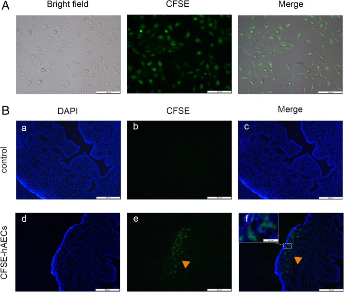
Fig. 3.
hAECs migrated to the injured uterine in vivo. A hAECs were labeled with CFSE before transplantation into the mice. The expression rate of green fluorescence staining was 100%. Scale bar = 100 μm. B According to immunostaining, CFSE-labeled hAECs (arrowheads) engrafted in the injured uterine at 3 days after hAEC transplantation, while no fluorescence-stained cells were detected in IUA mice without hAEC transplantation. a–f, scale bar = 200 μm; inset, scale bar = 25 μm

