Fig. 7.
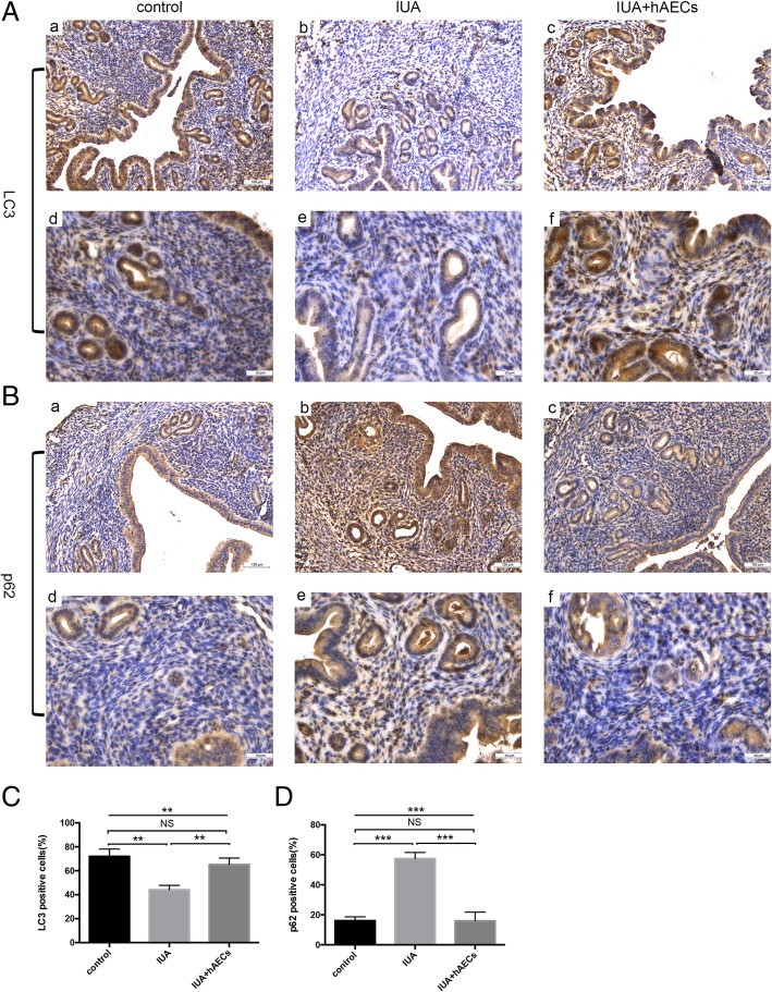
hAECs rescued impaired autophagy in the endometrium. A According to IHC staining, the number of LC3-positive cells was reduced in the IUA group and increased in the hAEC-treated group. B The number of p62-positive cells was increased in the IUA group and reduced in the hAEC-treated group. C, D Semi-quantification of LC3 and p62 expression in murine endometrium was calculated as the percentage of positive cells per field (*p < 0.05; **p < 0.01; ***p < 0.001; NS, p ≥ 0.05). a–c, scale bar = 100 μm; d–f, scale bar = 50 μm

