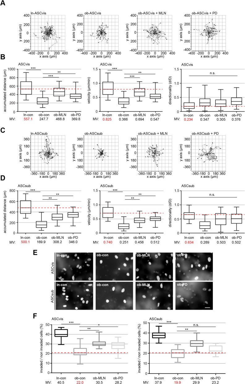
Fig. 3.
The rescue of primary cilia restores significantly the motility and invasion capacity of visceral and subcutaneous obese ASCs. a–d Analysis of cell motility of lean, obese, and obese ASCs treated with MLN or PD. Representative trajectories are depicted for individual cells (a and c, n = 30 cells in each group). The accumulated distance (left), the velocity (middle), and the directionality (right) are evaluated for indicated ASC as shown in the box plots (n = 90 cells pooled from three experiments). e Representatives of invaded visceral and subcutaneous ASCs (ln-con, ob-con, ob-MLN, and ob-PD) stained with DAPI. Scale bar, 30 μm. f Quantification of invaded ASCs. Percentage of invaded cells in comparison with the whole cell count. The results are presented as median ± min/max whiskers in box plots (visceral, left; subcutaneous, right) based on three independent experiments. An unpaired Mann-Whitney U test was used for statistical evaluation. ∗p < 0.05, ∗∗p < 0.01, ∗∗∗p < 0.001

