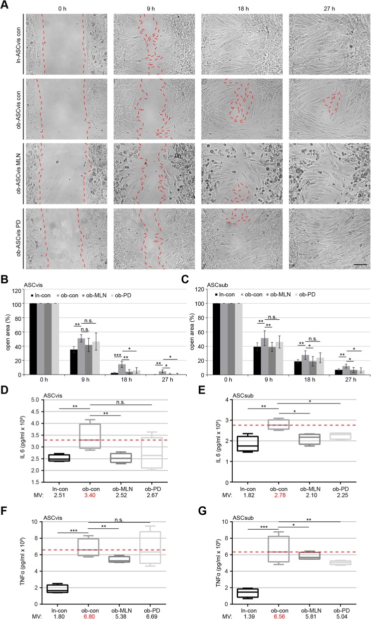
Fig. 4.
Reduced inflammatory cytokine secretion (IL6 and TNFα) and improved migration of obese ASCs after inhibitor treatment. a Wound healing/migration assays were performed with visceral and subcutaneous ASCs (ln-con, ob-con, ob-MLN, and ob-PD), and images were taken at indicated time points to document the migration front closure. Representatives are shown. Red dashed line depicts the migration front. Scale, 150 μm. b, c Quantification of the open area between both migration fronts at various time points is indicated (n = 5 visual fields of 1350 × 1800 μm2 for each condition). The cell-free area of each individual condition at 0 h was assigned as 100%. The results are based on three independent experiments with ASCs from three different donors (obese and lean) and presented as mean ± SEM. d–g 72-h supernatants of visceral (d, f) and subcutaneous (e, g) ASCs after 24-h pretreatment with indicated inhibitors were collected for the evaluation of IL6 (d, e) and TNFα (f, g). The results are from three experiments and presented as median ± min/max whiskers in box plots. Student’s t test was used for statistical evaluation for (b–g). ∗p < 0.05, ∗∗p < 0.01, ∗∗∗p < 0.001

