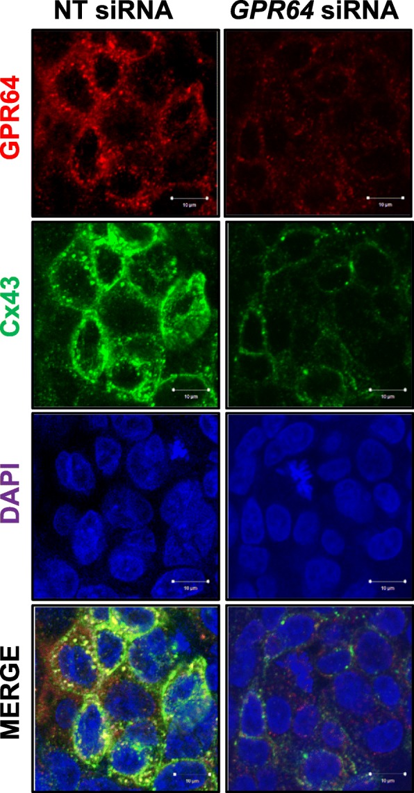
Fig. 6.

The colocalization of GPR64 with Cx43 in Ishikawa cells. The colocalization of GPR64 (red) and Cx43 (green) were analyzed in Ishikawa cells transfected with NT siRNA or GPR64 siRNA by fluorescence microscopy. GPR64 overlaps with Cx43, but its colocalization was affected by reduction of GPR64. Nuclei were counterstained with DAPI staining
