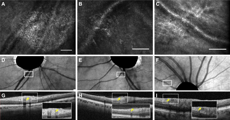
Figure 2.
Granular membranes on multimodal imaging. Scale bars: 100 μm. (A–C) Adaptive optics scanning laser ophthalmoscopy (AOSLO) en face images of granular membranes. (D–F) The corresponding location of the AOSLO membranes highlighted in white boxes on en face optical coherence tomography (OCT). The OCT images show masked optic nerve heads as automated by the machine. (G–I) The OCT B-scans in the corresponding region of interest. The cross sections in (G, H) with their insets illustrate subtle hyperreflective features above the retina (yellow arrow), while in (I) there is a suggestion of focal thickening of the internal limiting membrane (yellow arrow).

