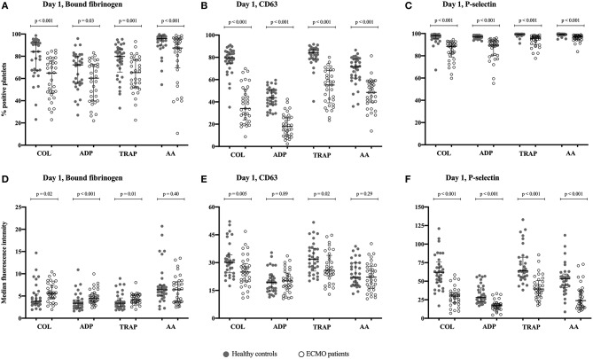
Figure 3.
Illustrates the degree of platelet activation in response to stimulation by four different agonists. The degree of activation can be measured as a change in the expression of the activation-dependent platelet surface markers bound-fibrinogen, CD63 and P-selectin on day 1 of support with extracorporeal membrane oxygenation (ECMO). The results are compared with healthy controls, n = 33. (A–C) The percentage of activated platelets in response to stimulation by an agonist; (D–F) The median fluorescence intensity (MFI) of the activated platelets following stimulation by an agonist. Agonists used to activate the platelets: COL, Collagen-related peptide; ADP, adenosine diphosphate; TRAP, thrombin receptor activating peptide-6; and AA, arachidonic acid. The bars indicate median and interquartile range.

