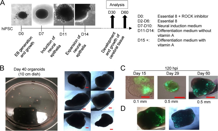
FIG 5.
Three-dimensional cortical organoid generation and infection by HCMV. (A) Multicellular organoids were generated over a time course of 30 to 60 days, with the developmental steps and culture conditions indicated during each phase of the differentiation process. Representative bright-field images of early stage organoids are shown. (B) Image of day 40 organoids prepared in a 10-cm culture dish and later moved to individual wells of a multiwell plate showing uniformity of size. Scale bar, 0.2 mm. (C) Day 15, 29, and 60 organoids were infected using HCMV TB40/E-eGFP based on an approximated area at 1 IU/μm2 in individual culture wells, and bright-field and fluorescent images obtained at 120 hpi. (D) Organoids prepared separately from those used in the experiment shown in panel B were infected and evaluated for GFP fluorescence at 120 hpi. D, day; ROCK, rho-associated coiled-coil protein kinase; hiPSC, human iPSC.

