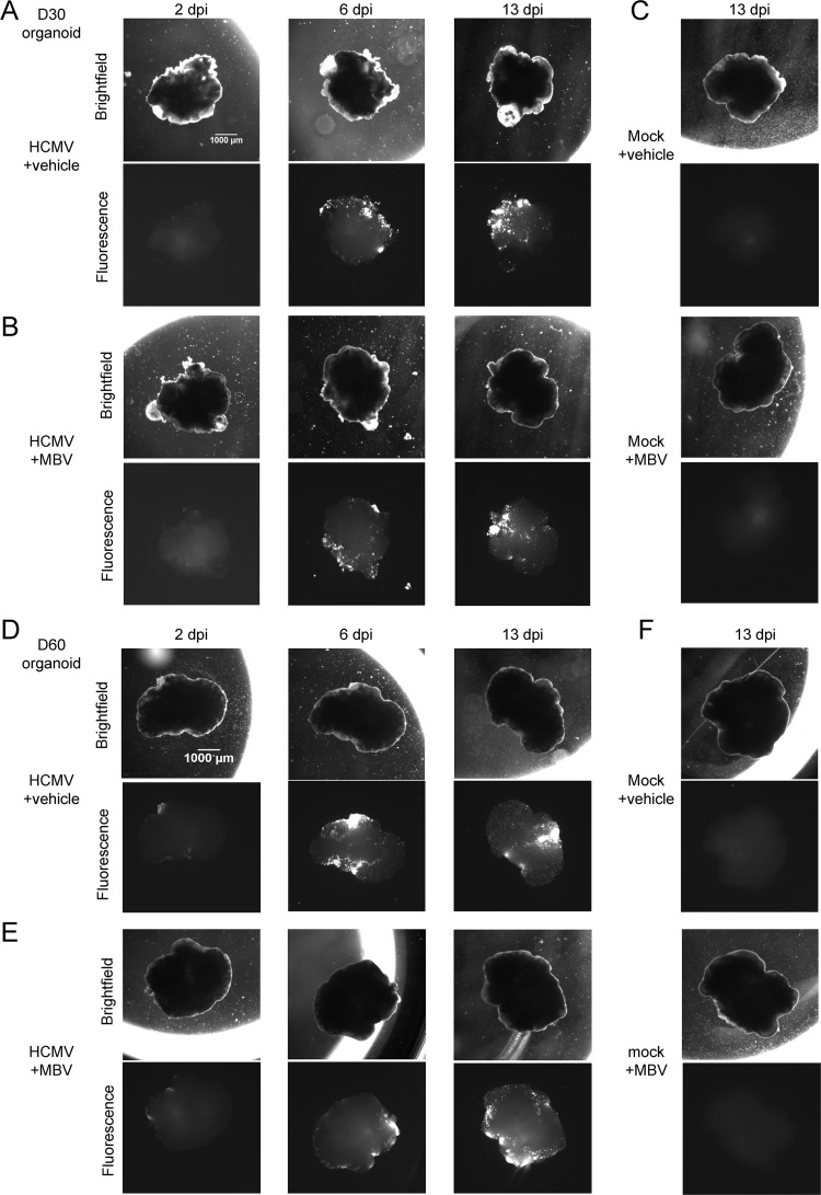
FIG 6.
HCMV infection spread in cortical organoids regardless of MBV cotreatment. (A to C) Representative bright-field and fluorescent images of day 30 (D30) organoids infected with HCMV TB40/E-eGFP at a multiplicity of 1 IU/μm2 between 2 and 13 dpi and treated with vehicle or 10 μM MBV or mock infected and treated as indicated. (D to F) Representative bright-field and fluorescent images of day 60 organoids infected with HCMV TB40/E-eGFP at a multiplicity of 1 IU/μm2 between 2 and 13 dpi and treated with vehicle or 10 μM MBV or mock infected and treated as indicated. Images were obtained using a 1× objective at the indicated times.

