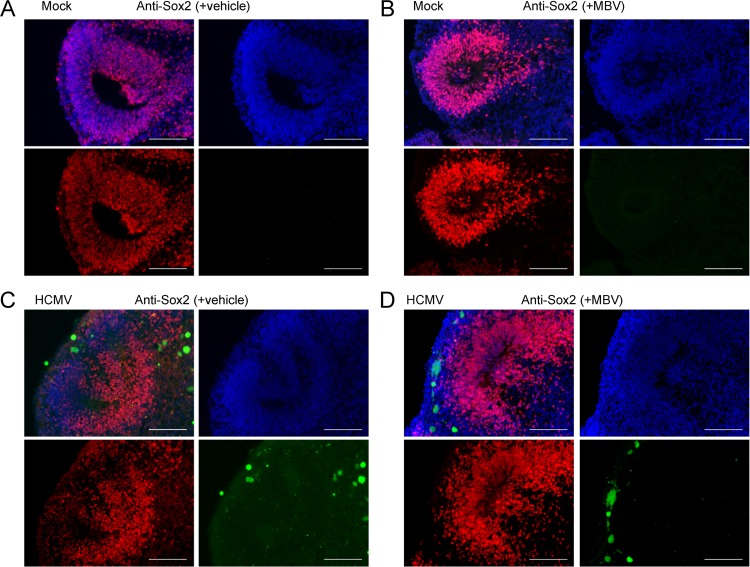
FIG 8.
HCMV-infected organoids show disrupted rosette formation and Sox2 expression patterns. Representative immunofluorescent images of cryosectioned day 30 organoids stained at 14 dpi for Sox2 expression (red) and with 4′,6′-diamidino-2-phenylindole (blue) following mock infection with vehicle (A), mock infection with 10 μM MBV (B), HCMV TB40/E-eGFP infection (green) with vehicle (C), and HCMV TB40/E-eGFP infection with MBV (D). No GFP fluorescence was observed under mock conditions. Scale bar, 100 μm.

