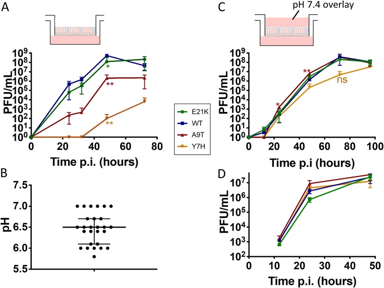
FIG 3.
Replicative ability in primary human airway epithelial cells cultured at the air-liquid interface. Primary human airway epithelial (pHAE) cells (A and C) or MDCK cells (D) were infected with each virus at low MOI, in triplicate. Time points taken at intervals postinfection were titrated by plaque assay. (C) A liquid overlay buffered to pH 7.4 was maintained on the apical surface. One-way ANOVA with Tukey’s posttest was used to compare WT virus to the other viruses. *, P < 0.05, **, P < 0.01; ***, P < 0.001; ns, not significant. Experiments are representative of the results from at least two biological replicates. (B) The pH of apical washes from 27 HAE cultures was tested using an unbuffered 0.9% saline (adjusted to pH 7.4) wash. The medians and interquartile ranges are shown.

