Figure 4.
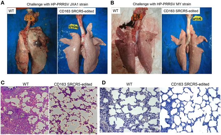
CD163 SRCR5-edited pigs exhibit normal histopathology after PRRSV challenge. Lungs were isolated from CD163 SRCR5-edited and WT animals on day 42 post PRRSV challenge. Pathological changes of lung lesions were observed and assessed using visual examination, Hematoxylin and Eosin (H&E) staining and immunohistochemistry. (A,B) Photographs of the dorsal side of lungs from CD163 SRCR5-edited and WT animals challenged with PRRSV JXA1 (A) and MY (B) strains. (C) Lung paraffin sections were stained with H&E (scale bar, 100 μm). (D) Immunohistochemistry analysis of the PRRSV antigen (brown) in lung paraffin sections (scale bar, 50 μm). The macrophages stain intensely dark brown due to the presence of the PRRSV antigen.

