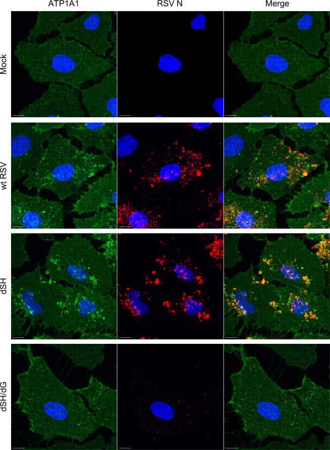
Fig 4. RSV G is required for ATP1A1 clustering.
A549 cells were inoculated with wt RSV, rgRSV-dSH or rgRSV-dSH/dG (MOI = 10 PFU/cell) and incubated for 5 h. Cells were fixed with 4% PFA and subjected to immunofluorescence staining, as described for Fig 3. ATP1A1 (green) was detected by an anti-ATP1A1 rabbit MAb (ab76020) and AF568-conjugated donkey anti-rabbit secondary antibody. RSV N (red) was detected by an anti-RSV N mouse MAb (ab94806) and an AF647-conjugated donkey anti-mouse secondary antibody. The cell nuclei were stained with DAPI (blue). Images (z-stacks) were acquired on a Leica SP8 confocal microscope, with a 63x objective (NA 1.4) and a zoom of 3.0. Scale bar 10 μm.

