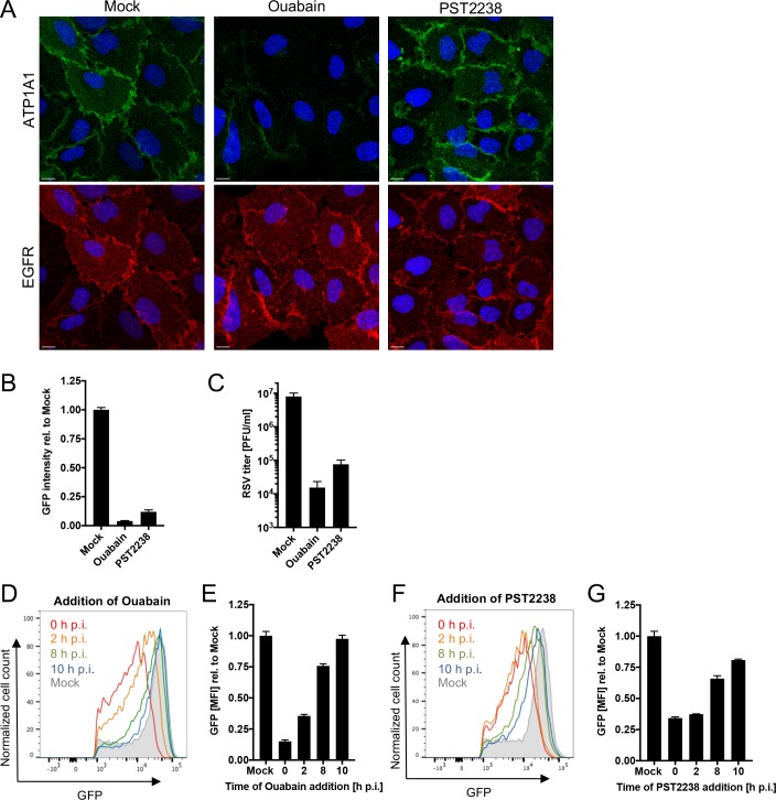
Fig 5. Effect of ouabain and PST2238 on RSV infection.
(A) ATP1A1 and EGFR expression in A549 cells treated with Ouabain or PST2238. Uninfected A549 cells were treated for 24 h with either 25 nM ouabain or 20 μM PST2238 and subjected to an immunofluorescence staining. ATP1A1 (green) was detected by an anti-ATP1A1 rabbit MAb (ab76020) and an AF488-conjugated donkey anti-rabbit secondary antibody. EGFR (red) was detected by an anti-EGFR rat MAb (ab231) and an AF647-conjugated goat anti-rat secondary antibody. The cell nuclei were stained with DAPI (blue). Scale bar 10 μm. (B and C) Inhibitory effect on RSV infection. A549 cells pre-treated for 16 h with either 25 nM ouabain or 20 μM PST2238 were inoculated with RSV-GFP (MOI = 1 PFU/cell). Cells were incubated for 17 h and infectivity was quantified by: (B) GFP signal of the total well, scanned by an ELISA reader and reported relative to mock-treated infected cells as 1.0; and (C) virus titer determined by plaque titration on Vero cells 24 h p.i. (D—G) Time-of-drug-addition experiment. A549 cells infected with RSV-GFP (MOI = 3 PFU/cell) were changed to medium containing 25 nM ouabain (D, E) or 20 μM PST2238 (F, G) at the indicated times post infection. Cells were harvested 24 h p.i. and GFP intensity was quantified by flow cytometry of live, single, GFP+ cells. The MFI of GFP+ cells was quantified and expressed relative to mock-treated, RSV-infected cells (E, G).

