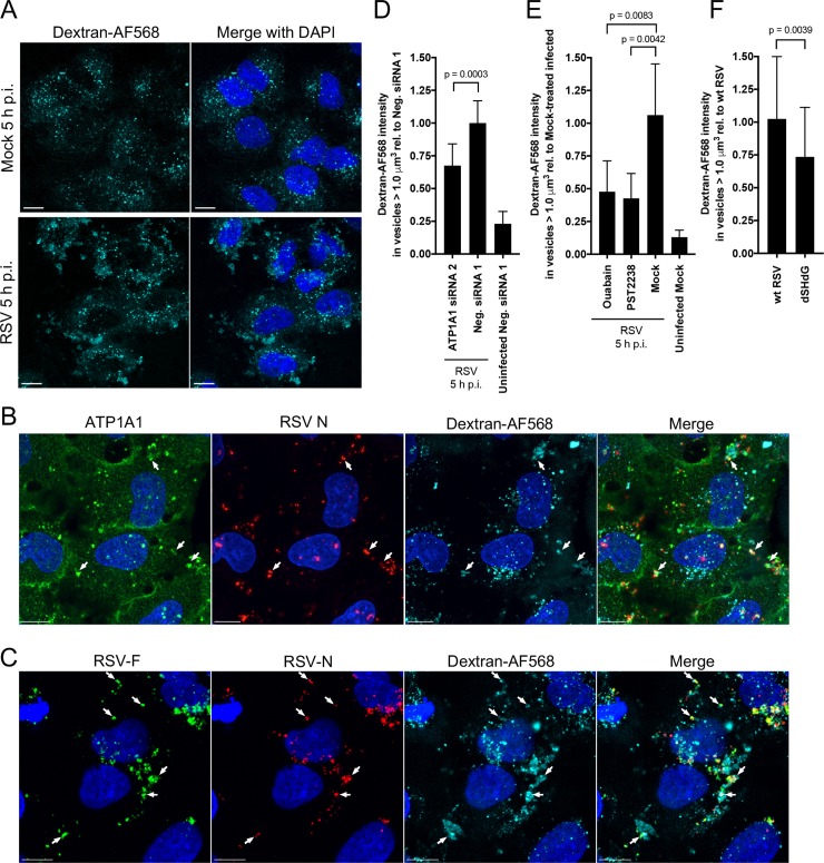
Fig 9. RSV induces and is taken up by ATP1A1-dependent macropinocytosis, which can be blocked by ouabain or PST2238.
Macropinocytosis was assayed by monitoring the uptake of dextran (10,000 MW) conjugated to AF568 (dextran-AF568). All incubations with dextran-AF568 were preceded by serum-starvation for 16 h. (A) RSV induces macropinocytosis. A549 cells were mock-infected or infected with wt RSV (MOI = 5 PFU/cell) in medium containing dextran-AF568 (cyan). At 5 h p.i., cells were fixed with 4% PFA and nuclei counterstained with DAPI (blue), and imaged on a Leica SP5 confocal microscope with a 40x Objective NA 1.3 and 2.0x zoom. (B) Co-localization of ATP1A1, RSV N, and dextran-AF568 in RSV-infected A549 cells. Cells were infected with RSV in the presence of dextran-AF568 as described above, incubated for 5 h, fixed with 4% PFA, permeabilized with 0.1% Triton X-100, subjected to immunofluorescence staining with an anti-ATP1A1 rabbit MAb (ab76020) and an anti-RSV-N mouse MAb (ab94806), followed by AF488-conjugated goat anti-rabbit and AF647-conjugated goat anti-mouse secondary antibodies. Z-stacks were acquired on Leica SP8 confocal microscope with 63x objective, NA 1.4 and 3.0x zoom. Arrows indicate co-localization of ATP1A1 (green) and RSV N (red) in dextran-AF568-positive (cyan) vesicles. (C) Co-localization of RSV F and RSV N with dextran-AF568 in RSV-infected A549 cells. Cells were infected with RSV in the presence of dextran-AF568, incubated for 5 h, fixed, and permeabilized as described in B. The cells were then subjected to immunostaining: RSV F was detected with AF488-conjugated anti-RSV F MAb #1129 [72], and RSV N was detected with an allophycocyanin (APC)-conjugated anti-RSV N MAb (NB100-64752APC, Novus Biologicals, LLC). Image acquisition and analysis were performed as described above for B. Arrows indicate RSV F (green) and RSV N (red) in dextran-AF568-positive (cyan) vesicles. All scale bars are 10 μm. (D–F) Quantification of dextran-AF568 uptake during RSV infection. (D) A549 cells were transfected with ATP1A1 siRNA2 or Neg. siRNA 1, incubated for 48 h p.t., and inoculated with wt RSV in dextran-AF568-containing medium, or (E) A549 cells were pre-treated with ouabain or PST2238 for 16 h and inoculated with wt RSV in dextran-AF568-containing medium, or (F) A549 cells were infected with wt RSV or rgRSV dSH/dG in dextran-AF568-containing medium. For all treatments (D-F) cells were fixed 5 h p.i., counterstained with DAPI and z-stacks were acquired on a Leica SP8 confocal microscope with 63x objective NA 1.4, 1.0x zoom. For each treatment, the uptake of dextran-AF568 in vesicles greater than 1.0 μm3 was quantified as described in detail in the Materials and Methods section. Mean values are reported relative to RSV-infected cells transfected with Neg. siRNA 1 (D), or mock-treated infected cells (E), or wt RSV-infected cells (F). Error bars indicate the standard deviation of at least three independent experiments. The statistical significance of difference was determined for (D) and (E) by one-way analysis of variance with Tukey’s multiple comparison post-test and for (F) by a two-tailed unpaired t-test. P-values are shown for each comparison.

