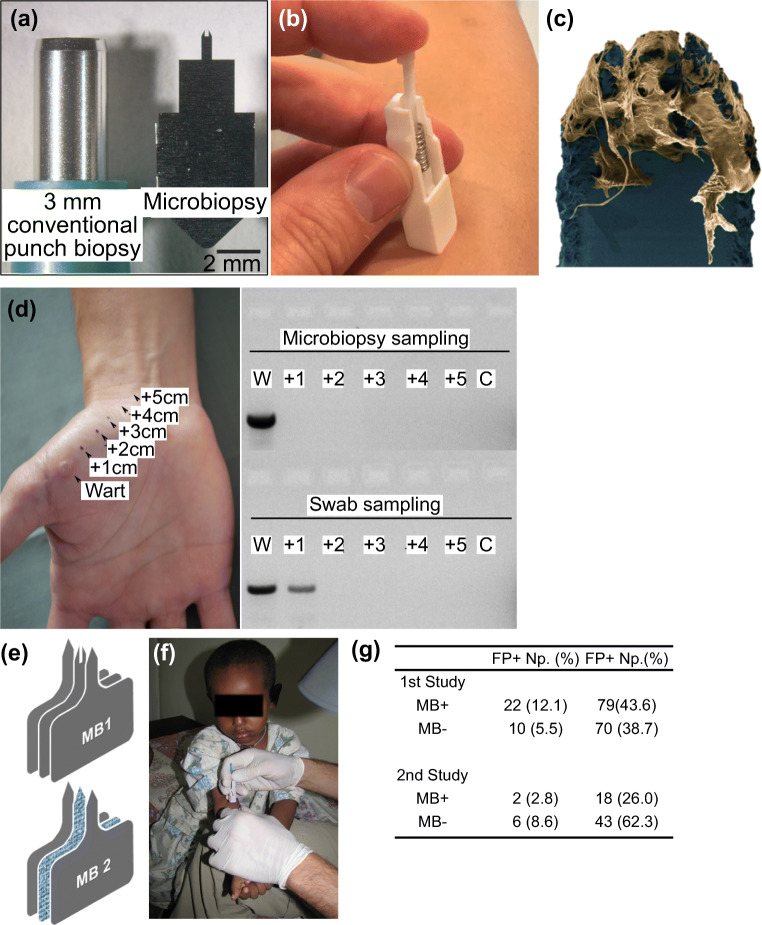
Fig. 7.
Microneedle-based microbiopsies were used in skin and blood microsampling. a) A side-by-side comparison between conventional punch biopsy and the skin microbiopsy. b) The spring-loaded applicator used in microbiopsy application. c) After application, the microneedle captured pieces of skin tissue in the channel. d) Spatial detection of HPV by sampling cutaneous warts. Skin microbiopsy provided a more accurate spatial detection as demonstrated by DNA gel electrophoresis. e) Designs of skin (top) and absorbent microbiopsies (bottom). The main difference between the two designs was the middle absorbent layer. f) The absorbent microbiopsy was used to sample patients with Leishmaniasis in rural areas. g) The PCR data suggested the absorbent microbiopsy was able to detect Leishmaniasis more accurately than the finger-prick method. Pictures retrieved from Lin et al. 2013, Tom et al. 2016 and Kirstein et al. 2017. All figures are under a Creative Commons Attribution 2.0. Full terms at http://creativecommons.org/licenses/by/2.0

