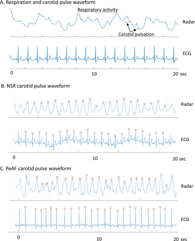
Figure 4.
Representative radar signal waveforms of respiratory activity and the carotid pulses. After the removal of clutter, the signal waveform was composed of large cyclic waves from respiratory activity and small but recognizable peaks from carotid pulsation (A). After high- and low-pass filters were applied, and the waveform was smoothed over 3 data points, the peak points on the carotid radar pulse waveform appeared to be well-correlated with R waves on ECG in both patients with NSR (B) and those with PeAF (C). The red circles at each peak in the radar waveform indicate the systolic phase of the carotid pulsation corresponding to each ECG R wave.

