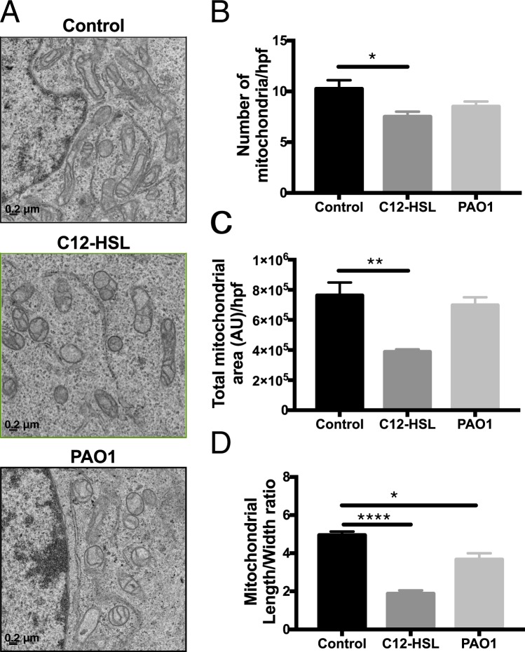
Figure 1.
P. aeruginosa QS molecules disrupt mitochondrial morphology in bronchial epithelial cells. BEAS-2B cells were treated with vehicle control (DMSO), 100 μM 3-oxo-C12-HSL (C12-HSL), or infected with PAO1 (MOI 20) for 6 hours. Cells were then fixed and processed for electron microscopy (EM) analysis. (A) representative high-powered field (hpf) images of treatment groups, 15,000x magnification, scale 0.2 μm. (B) number of mitochondria per hpf. (C) Total mitochondrial area per hpf. (D) Mitochondrial length to width ratio. 3-oxo-C12-HSL and PAO1 disrupt mitochondrial morphologic parameters. Results are mean ± SEM. *P < 0.05, **p < 0.01, ****p < 0.0001 all vs. control. One-way ANOVA with Tukey’s multiple comparisons test used for statistical analysis.

