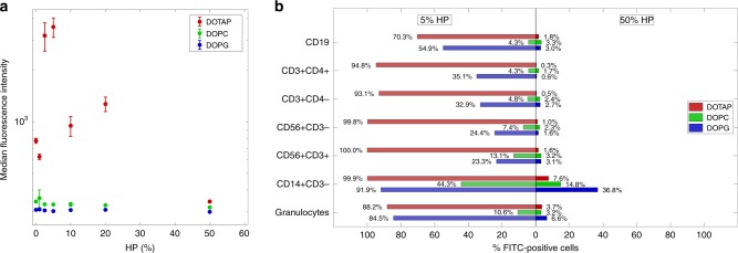
Fig. 4.
Sequestration of liposome–protein complexes by immune cells in vitro. a Cellular uptake of fluorescently labeled liposome–protein complexes by human monocyte THP1 cells via flow cytometry: DOTAP (red), DOPC (green), and DOPG (blue). Complexes were prepared at several human plasma (HP) concentrations: 0 (pristine liposomes), 1%, 2.5%, 5%, 10%, 20%, and 50%. Each value is the average of triplicate samples ± standard deviation within a single experiment. b Leukocyte uptake of liposome–protein complexes via flow cytometry: DOTAP (red), DOPC (green), and DOPG (blue). Complexes were prepared at low (5%) and high (50%) human plasma (HP) concentrations. Each value is the average of duplicate samples ± standard deviation within a single experiment. The fluorescence of internalized liposomes was measured as the percentage of FITC-positive cells by gating on distinct leukocyte subpopulations as indicated. The gating strategy was obtained as shown in Supplementary Fig. 3

