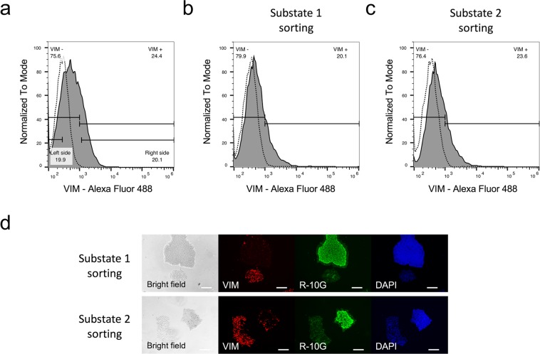
Figure 2.
Flow cytometry and immunohistochemical analyses in the H9 cell cultured on Matrigel. (a–c) Histograms of the H9 cells incubated with anti-vimentin antibodies (solid line) or isotype control (dash line). The substate 1 and substate 2 cells were separated from approximately 20% on the left and right side of vimentin signals, respectively (a). Flow cytometry analyses of substate 1 and substate 2 passaged one time after sorting (b,c). Data are representative of three independent experiments. (d) Immunohistochemistry with no permeabilisation using antibodies for a pluripotency marker (R-10G epitope) and a mesenchymal marker (vimentin [VIM]). Nuclei were counterstained with DAPI. The scale bar represents 200 µm.

