
Figure 3.
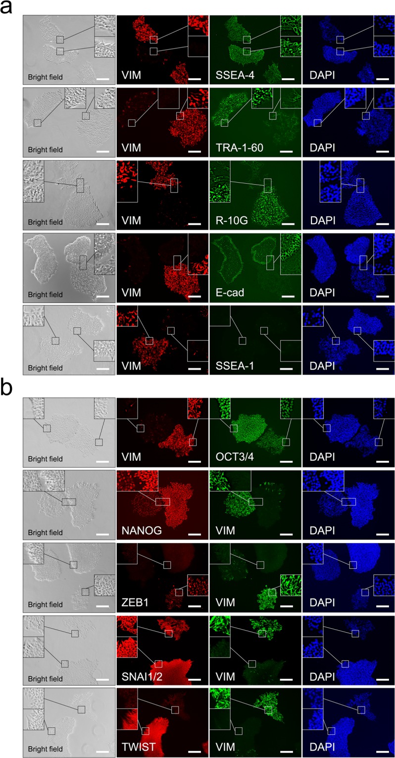
Immunohistochemical analysis of pluripotency, differentiation, and epithelial-to-mesenchymal transition markers in H9 cells cultured on Matrigel. (a) Immunohistochemistry with no permeabilisation using antibodies for vimentin (VIM), SSEA-4, TRA-1-60, R-10G, E-cadherin (E-cad), and SSEA-1. (b) Immunohistochemistry with permeabilisation using antibodies for VIM, OCT3/4, NANOG, ZEB1, SNAI1/2, and TWIST. Nuclei were counterstained with DAPI. The inset shows a 3 × enlargement of the figure. The scale bar represents 200 µm.