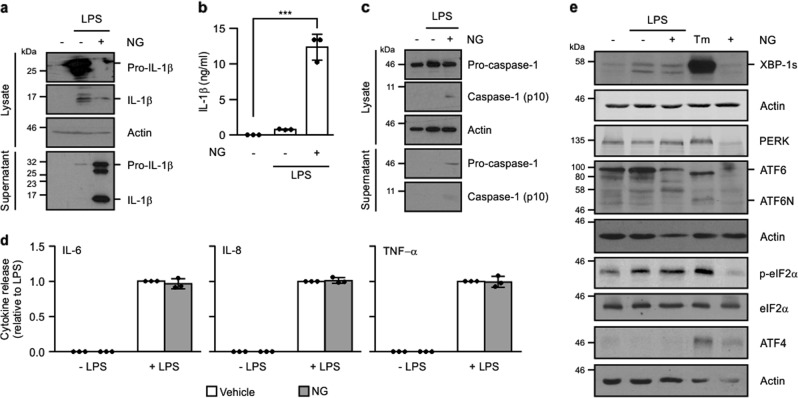
Fig. 1. IRE1α-XBP1 axis of the UPR is selectively activated upon TLR4 stimulation.
THP-1 cells were primed with either 1 μg/ml LPS alone for 24 h or 1 μg/ml LPS for 24 h followed by addition of 10 μM nigericin (NG) for 45 min. a Processing of pro-IL-1β was analysed in cell lysates and conditioned medium by immunoblotting for full-length pro-IL-1β and processed p17 IL-1β. b Levels of IL-1β were quantified in conditioned medium from untreated, LPS, LPS and NG-treated THP-1 cells by ELISA (n = 3). c Processing of caspase-1 was analysed in cell lysates and conditioned medium by immunoblotting for pro-caspase-1 and processed p10 caspase-1. d IL-6, IL-8, and TNF-α levels were quantified in conditioned medium from untreated, LPS, LPS and NG, and NG-only treated THP-1 cells by ELISA (n = 3). e UPR markers XBP1s, PERK, ATF6, phospho-eIF2α, total eIF2α, and ATF4 were analysed by immunoblotting in THP-1 post treatment with LPS alone or LPS and NG. Tunicamycin (Tm)-treated THP-1 cells served as a positive control for the UPR activation. Actin was used as a loading control. ***P < 0.001 based on a Student’s t test. Error bars represent SD.

