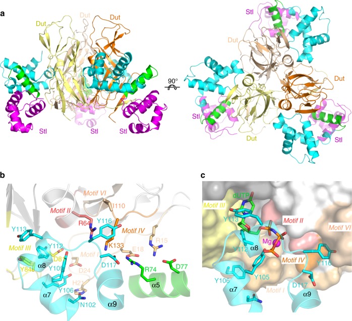
Fig. 3.
Crystal structure of the BovI-StlN-ter-Dut ϕ11 complex. a Structure of the complex between the trimeric Dut from phage ϕ11 (protomers coloured in different tones of yellow-orange) and three molecules of BovI-StlN-ter (protomers coloured as in Fig. 1b). Two orthogonal views are shown. b Close-up view of the interaction of a protomer of BovI-StlN-ter with the Dutϕ11 trimer. Notice that only two of the three protomers of Dutϕ11 contact each Stl molecule. Dut catalytic motifs are highlighted in different colours and labelled. The residues involved in interactions are shown as sticks, labelled and coloured by atom type with the carbons in the same colour as the corresponding molecule. For clarity, only the structural elements of BovI-StlN-ter involved in the interaction are shown and labelled. c Stl mimics the interactions of the dUTP substrate with Dutϕ11. The substrate dUTP was placed in the active centre of Dutϕ11 in complex with BovI-StlN-ter by superimposing the structure of Dutϕ11-dUTP (PDB 4GV8) and is shown as sticks with carbon atoms in green. The Mg ion chelated by the dUTP is shown as a magenta sphere. Dutϕ11 is represented in surface and BovI-StlN-ter in cartoon and sticks. Structural elements and BovI-StlN-ter interacting residues are labelled as well as conserved catalytic motifs of Duts and coloured as in b. The omit electron density map is shown in Supplementary Fig. 13. Source data are provided as a Source Data file

