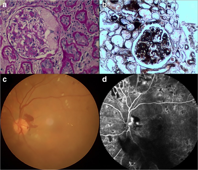
Fig. 1.
Example of diabetic retinopathy and nephropathy (DRN) seen on renal biopsy and retinal fundus. Hematoxylin-eosin staining (a) and cyclic acid-silver-methylamine staining (b) of renal tissue showed severe dilation of mesangial matrix and tuberous sclerosis of glomerulus. Funduscopy (c) showed that the retina had diffuse microaneurysms and patches of hemorrhage. The fluorescein fundus angiography (d) showed a significant increase in the number of retinal microangiomas

