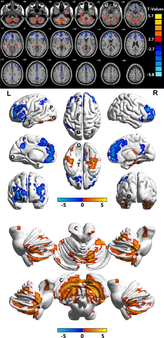
Fig. 2.
Marked differences in spontaneous brain activity in the DRN group compared with HCs. Notes: The different brain regions were observed in the bilateral cerebellum posterior/anterior lobe, left inferior temporal gyrus, bilateral medial frontal gyrus, right superior temporal gyrus, right middle frontal gyrus, left middle/inferior frontal gyrus, bilateral precuneus, and left inferior parietal lobule in the DRN group. The red areas denote higher ALFF brain regions, and the blue areas denote lower ALFF brain regions. ALFF, amplitude of low-frequency fluctuation; HCs, healthy controls; L, left; R, right; B, bilateral; T, temporal lobe; F, frontal lobe; O, occipital lobe; C, cerebellum; P, parietal lobe

