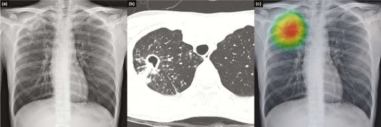
Figure 3.
Representative case from the observer performance test. Chest radiograph of a 25-year-old woman shows a cavitary mass with multiple satellite nodules in the right upper lung field (a), which corresponded well with computed tomography images. These radiologic findings are typical for active pulmonary tuberculosis (b). Deep learning–based automatic detection algorithm provided a probability value of 0.9663 for active pulmonary tuberculosis in this case, and the classification activation map correctly localized the lesion in the right upper lung field (c).

