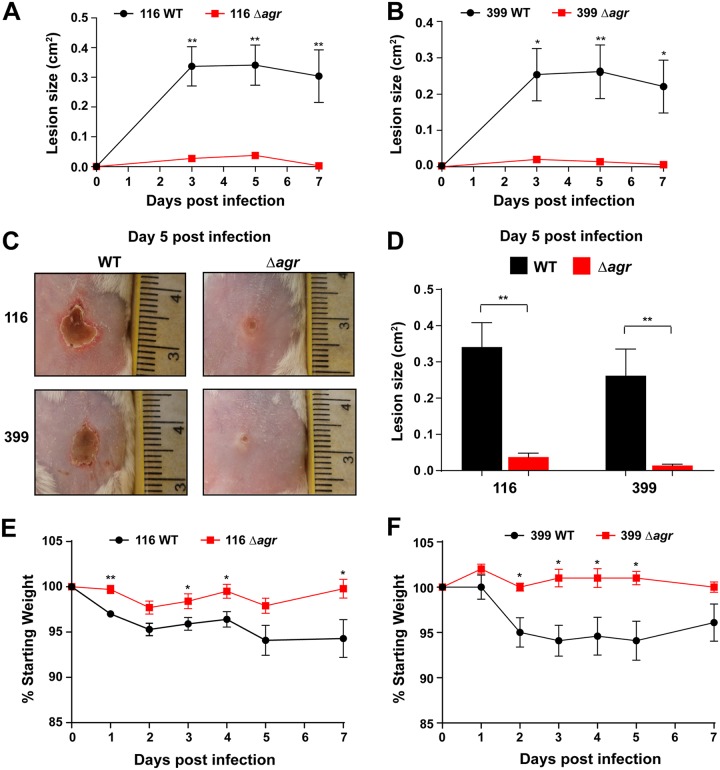
FIG 4.
Role of agr in USA100 skin infections. (A, B) Comparison of lesion size for mice inoculated with the strain 116 (A) and 399 (B) WT and Δagr mutant strain pairs over 1 week (n = 5 for each group). (C) Photographs of ventral skin lesions 5 days after inoculation with WT and Δagr mutant strains. Note the smaller areas of dermal necrosis resulting from infection with the Δagr mutants. (D) Comparison of lesion size between WT and Δagr mutant isolates 5 days after intradermal injection of mice (n = 5 for each group). (E, F) Comparison of weight loss (expressed as a percentage of the starting weight) in mice which underwent intradermal injections of the strain 116 (E) and 399 (F) WT and Δagr isolates. Statistical significance was determined using Student's t test. **, P < 0.01; *, P < 0.05.

