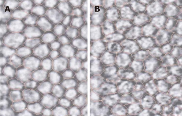
Figure 1.
Indirect specular microscopy images of corneal endothelium in normal eyes and eyes with primary open-angle glaucoma. A: Corneal endothelial cells showed regular hexagonal monolayer with clear cell borders in normal eyes; B: Endothelial cells showed irregular and indistinct cell borders with guttae like dark cells in patients with glaucoma.

