. 2018 Feb 6;2(4):724–750. doi: 10.1039/c7se00582b

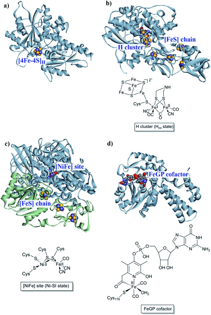
Fig. 1. [NiFe], [FeFe] and [Fe] hydrogenases. (a) The “apo” form of Chlamydomonas reinhardtii HydA1 ([FeFe] hydrogenase; PDB: ; 3LX4);24 (b) Clostridium pasteurianum CpI ([FeFe hydrogenase]; PDB: ; 4XDC);25 (c) Allochromatium vinosum [NiFe] hydrogenase (PDB: ; 3MYR);26 (d) Methanocaldococcus jannaschii [Fe] hydrogenase (PDB: ; 3DAG).27 Protein backbones are shown as solid ribbons and coloured by chain, and metal cofactors are shown as space-filling models and coloured by element (structures drawn using BIOVIA Discovery Studio Visualizer). A schematic representation of the active site of the different subclasses is shown below the respective enzyme structure. Heteroatom colour coding: S: yellow; Fe: indigo; Ni: dark blue; O: red; N: blue; P: orange.

Fig. 1