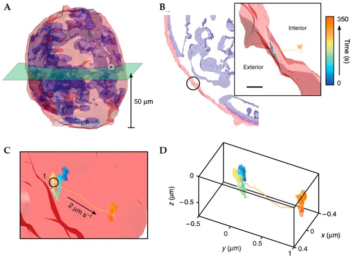
Figure 12.
Prior volumetric imaging combined with TSUNAMI-based tracking was used to acquire a volumetric image of the entire 100 µm diameter tumor spheroid around an internalizing particle. (A) 3D isocontour reconstruction of tumor spheroid with red plasma membrane and blue nucleus. Green section at 50 µm shows plane of EGFR internalization trajectory. (B) Isocontour model of the green slice in (A). Inset shows zoomed in view highlighting spheroid boundary and the placement of the trajectory as it transports into the spheroid. (C) Zoom in (B). (D) Isolated trajectory. Adapted from reference [92] under Creative Commons Attribution 4.0 International License.

