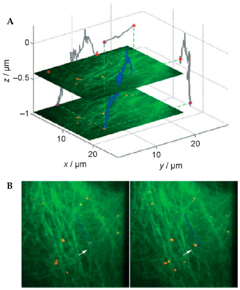
Figure 13.
The addition of simultaneous widefield imaging to orbital tracking allowed trajectories (blue) of artificial viruses (red) to be placed in the environmental context of their interactions with the cytoskeleton as visualized with eGFP-labeled tubulin (green). (A) Trajectory of an artificial virus shown in 3D space with 2D projections across each axis shown in gray. (B) Two frames in a time series of images with trajectory overlaid. In the left frame, the particle is moving laterally not because the virus itself is changing microtubules, but because the microtubule itself was moving. This observation could not have been confirmed without simultaneously acquired imaging. Reprinted from reference [84]. Copyright 2009 Wiley-VCH.

