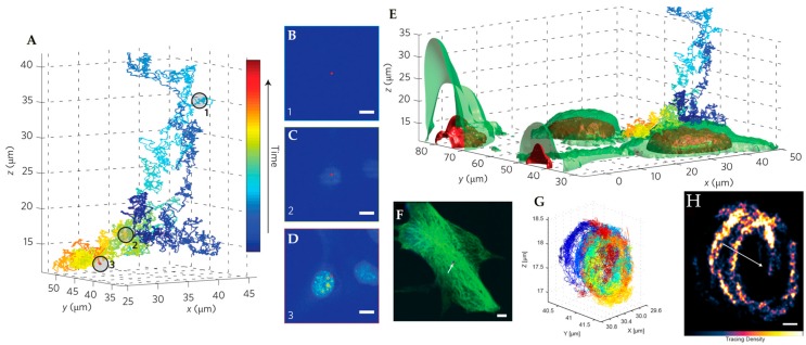
Figure 15.
3D Multi-Resolution Microscopy combines RT-3D-SPT with simultaneously acquired 2P-LSM images. (A) Isolated trajectory of QD-labeled nanoparticle probe. (B–D) Corresponding 2P-LSM sections acquired at depths indicated in circled portions of trajectory in (A). (E) Overlay of co-registered trajectory with 3D reconstruction of interpolated 2P-LSM sections gives cellular context to trajectory motion. Nuclear regions are highlighted in red and cell membrane surfaces are green. (F) 2P-LSM maximum intensity projection of internalized QD-labeled probe in a NIH-3T3 fibroblast cell. Arrow indicates position of tracked particle. (G) Ellipsoidal trajectory of macropinosomal membrane-bound probe showing multiple curved surfaces due to macropinosomal motion. (H) Structural tracing of the trajectory shown in (E). Arrow shows center of mass motion during trajectory. Adapted from reference [76] with permission of the author.

