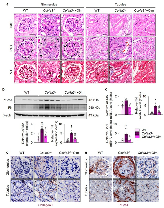
Figure 1.

Olmesartan ameliorates kidney fibrosis in Col4a3–/– mice. (a) Tissue morphology of kidney from WT, Col4a3–/–, and Col4a3-/-+Olm mice. Images from glomerulus (left) and tubulointerstitium (right) are presented. H&E, hematoxylin, and eosin staining; PAS, periodic acid–Schiff staining; MT, Masson’s trichrome staining. Note that, contrary to kidneys from the other groups, glomerular capillary lumens are barely patent (arrowheads) with shrinkage of mesangium (white asterisk) in Col4a3–/– mice. In Col4a3–/– mice, crescent are packing the Bowman’s space (arrows). Detachment of tubular epithelial cells from basement membrane (red arrowheads) and collagen deposits (white arrows) are also indicated. Scale bars, 50 μm. (b,c) Comparison of expression level for fibrosis markers determined by immunoblotting (b) and qPCR (c) from the kidney of WT, Col4a3–/–, and Col4a3-/-+Olm mice (n = 4 mice/group). β-actin was used as the endogenous control. (d,e) Representative images of immunohistochemical staining for collagen type 1 (d) and αSMA (d) in the kidney of WT, Col4a3–/–, and Col4a3-/-+Olm mice. Images from glomeruli (upper) and tubules (lower) are presented. Scale bars, 50 μm. * P < 0.05, ** P < 0.01 versus WT mice; # P < 0.05, ## P < 0.01 vs. Col4a3–/– mice by one-way ANOVA with Newman–Keuls multiple comparison test.
