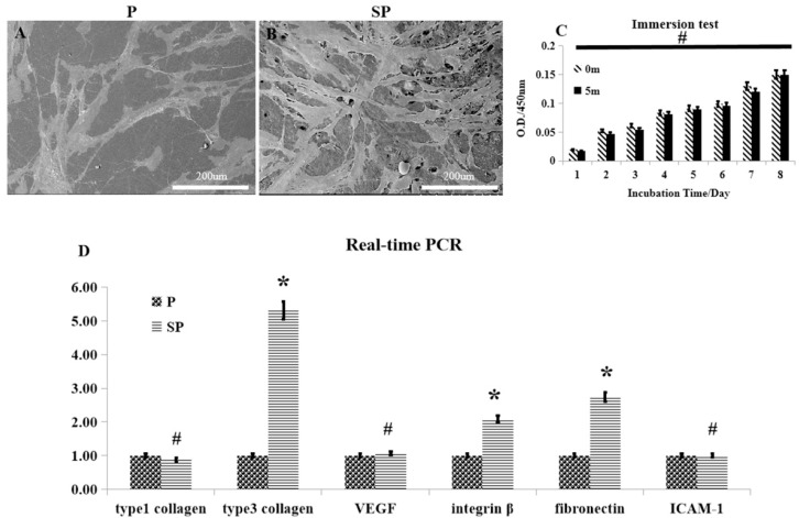
Figure 5.
The biological behavior of human skin fibroblasts (HSFs) on the surface of the peek materials. (A,B) SEM images of HSFs adhering on the peek specimens, and acid-etched peek specimens. (C) Comparison of the influence of P and SP for HSF adhesion. (D) RT-PCR of collagen I, collagen III, VEGF, integrin-β, ICAM-1, and GAPDH 5 days after spreading the HSFs on the specimens. * p < 0.05, # p > 0.05.

