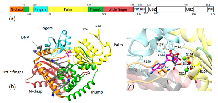
Figure 2.
(a) Diagram of the domains of human pol κ: In addition to the polymerase domains, PCNA-interacting peptide (PIP) regions, Rev1-interacting region (RIR), and ubiquitin-binding zinc finger (UBZ) domains are shown. (b) The structure of the polymerase domain of human pol κ with domains colored as in Figure 2a (PDB ID: 6CST) [23]. (c) Close-up view of the structure of pol κ highlighting the active site residues in contact with the incoming nucleotide. The catalytic residues D107, D198, and E199 are shown in yellow sticks with red oxygen atoms; others are shown as sticks colored by the domain as in Figure 2a,b. Metal ions are shown as green spheres.

