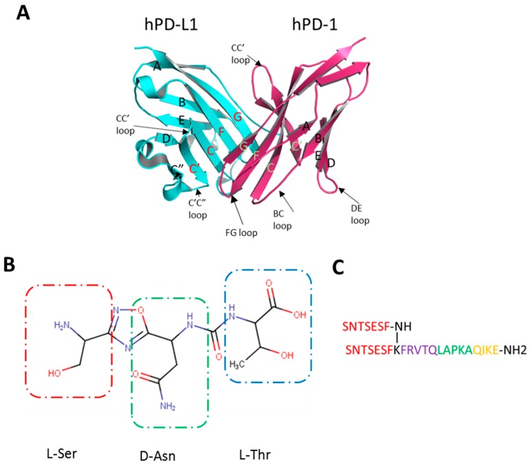
Figure 1.
(A) Structure of the hPD-1/hPD-L1 complex. hPD-L1 in cyan, hPD-1 in purple; The strands of IgV are shown with names. Strands involved in the protein-protein interaction (PPI) are coded in red for hPD-L1 and green for hPD-1 (adapted from [9]). (B) The putative structure of CA-170 with building blocks indicated by boxes. (C) Structure of the AUNP-12 peptide composed of 4 hPD-1 parts: 2x BC loop (SNTSESF) in red connected via lysine to the D strand (sequence FRVTQ), purple; the FG loop (sequence LAPKA) in green, and the G strand (sequence QIKE), orange.

