Figure 6.
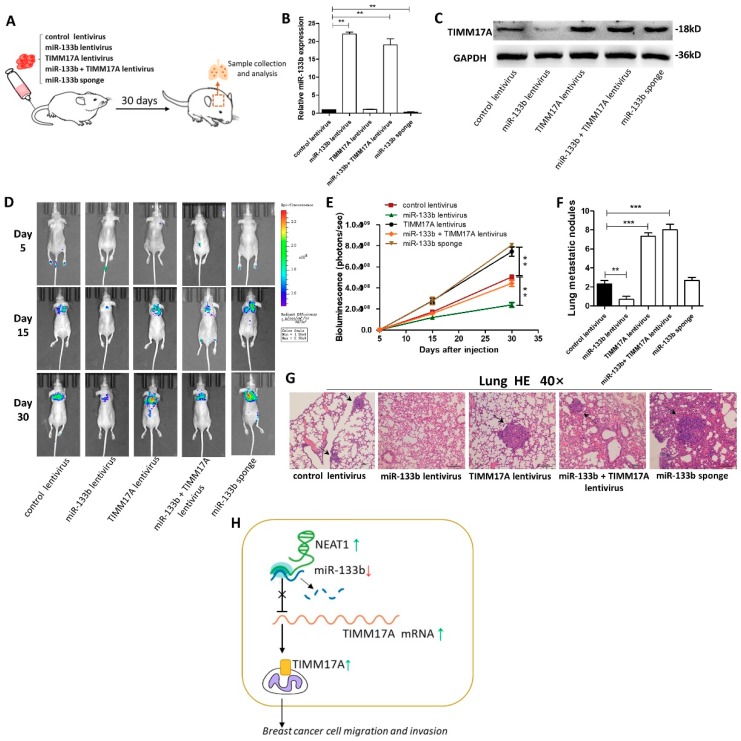
Effects of TIMM17A-targeted miR-133b on the lung and liver colonization of MDA-MB-231 cells xenografts in mice. (A) Experimental design: immunocompromised mice were injected through tail vein with MDA-MB-231 cells transfected with either the control lentivirus, miR-133b lentivirus, TIMM17A lentivirus, miR-133b lentivirus plus TIMM17A lentivirus, or miR-133b sponge. (B,C) miR-133b levels (B) and TIMM17A protein levels (C) in MDA-MB-231 cells transfected with either the control lentivirus, miR-133b lentivirus, TIMM17A lentivirus, miR-133b lentivirus plus TIMM17A lentivirus, or miR-133b sponge. (D,E) Representative BLI images (D) and quantitative analysis of the fluorescence intensities (E) of mice of five groups. The BLI was performed on days 5, 15, and 30 after injection. The intensity of BLI is represented by the color. (F,G) Numbers of metastatic nodules(F) and representative H&E-stained sections of lung tissues isolated from the intravenously injected mice. Black arrows indicate metastatic nodules. (H) A working model for the role of NEAT1-silenced miR-133b in breast cancer metastasis. During breast tumorigenesis, silencing of miR-133b mediated by NEAT1 facilitated migration and invasion of breast cancer cells through up-regulating the expression of TIMM17A. Scale bar, 200 μm. ** p < 0.01; *** p < 0.001.

