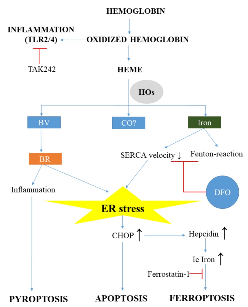
Figure 3.
Free hemoglobin and heme participates in the pathophysiology of brain damage. During brain hemorrhage, massive amounts of free hemoglobin and heme are released, resulting in inflammation, heme-, and ER stress. Oxidized hemoglobin directly triggers inflammation via toll-like receptor 2 and 4 (TLR2, TLR4). Heme oxygenases (HOs) in the brain are Janus-faced and might induce both protective and adverse effects. Although bilirubin (BR) is a natural antioxidant, it can induce inflammation, pyroptosis, or even apoptosis through ER stress in the brain. Heme-derived iron also triggers ER stress. Elevated intracellular iron (Ic Iron) derives from HO activity and the disturbed cellular iron metabolism through hepcidin induction, leading to ferroptosis. (SERCA: sarco/endoplasmic reticulum Ca2+-ATPase; DFO: desferrioxamine; CHOP: C/EBP homologous protein; CO: carbon monoxide; BV: biliverdin; TAK: Ethyl-(6R)-6-(N-(2-chloro-4-fluorophenyl)sulfamoyl)cyclohex-1-ene-1-carboxylate; TLR: Toll-like receptor

