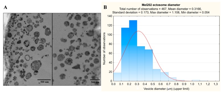
Figure 1.
TEM analysis of Mel202-derived ectosomes. Samples were fixed with 2.5% glutaraldehyde, postfixed in 1% osmium tetroxide, dehydrated by passing them through a graded ethanol series, and embedded in epoxy resin. Ultrathin sections were contrasted using uranyl acetate and lead citrate. The sections were viewed on a JEOL JEM 2100HT TEM at 80 kV (A). Subsequently, diameters of n = 467 ectosomes were measured with the use of ImageJ software (B).

