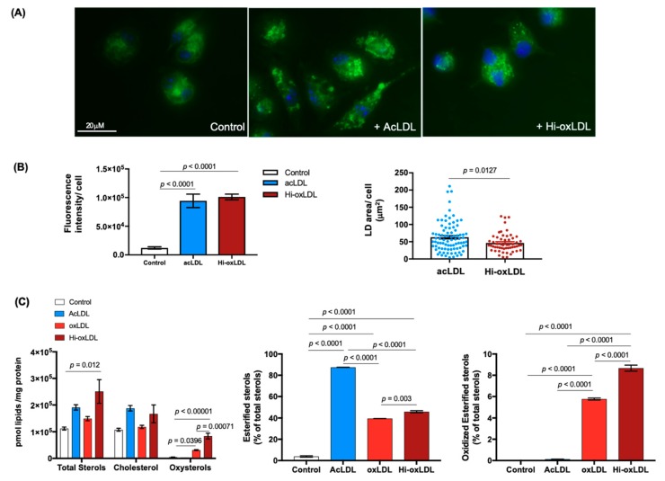
Figure 1.
Oxidized low-density lipoprotein (LDL) enriches oxysterols in macrophage foam cells. (A) Foam cell formation. Mouse peritoneal macrophages (MPMs) plated on a coverslip remained untreated (left) or were loaded with either acLDL (middle) or hi-oxLDL (right) for 24 h to generate low-density lipoprotein (LDs). LDs were visualized with Bodipy 483/503 staining (green) and nuclei were stained with DAPI (blue). (B) Total Bodipy fluorescence intensity per cell represents total lipid contents (left). Area of LD per cell was measured and presented in μm2/cell. n = 88 for acLDL and n = 55 for Hi-oxLDL-treated MPMs. (C) Sterol contents of foam cells treated with modified LDLs. MPMs were treated with acLDL for 24 h, and oxLDL or hi-oxLDL for 48 h. Detected sterols using mass-spectrometry (MS) was normalized by mg protein and shown as pmol of lipids over μg of protein. The data were presented as mean ± standard error of mean (SEM).

