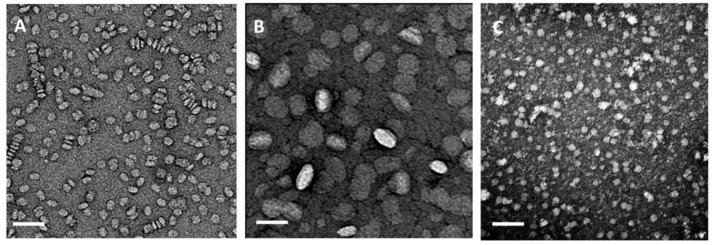
Figure 5.
Negative staining transmission electron microscopic (TEM) images of HDL particles. (A) rHDL particles reconstituted from dimyristoyl-phosphatidylcholine (DMPC) (8% cholesterol) and full length apo-AI at a molar ratio of 80:1 lipid:protein. The particles are about 10 nm in diameter and discoidal in shape. The circular shapes present nanodiscs viewed from the top. Likewise, stacked nanodiscs resembling “rouleaux” formations are visible. (B) rHDL particles assembled from DMPC (8% cholesterol) and apo-AI peptidomimetics (4F) at a molar ratio of 40:1 lipid to peptide. The nanoparticles are larger (about 25–30 nm) and seem to be ellipsoidal in shape; (C) HDL particles isolated from human plasma are spherical with particles sizes in the range from 8 nm to 14 nm (unpublished data). The bar represents 50 nm.

